This review presents the studies and the results achieved in the framework of the Virtual X-ray Reading – VXR project devoted to X-ray tomography for manuscripts digitization. The project started in 2014 at École Polytechnique Fédérale de Lausanne (EPFL, Switzerland) and was part of the ambitious Venice Time Machine project. VXR aimed to test the feasibility and to develop an alternate digitization technique for ancient manuscripts based on X-ray tomography. Research and technology made considerable progress in increasing the speed and the safety of the traditional digitization process of ancient collections, but, despite this, imaging of ancient, fragile, or un-opened documents remains a formidable challenge. Thanks to the high penetration of X-rays, the acquisition of a 3D – tomographic – volume is possible even without opening the document. The X-ray contrast necessary for the readability of the text is strictly connected to the chemical composition of the ancient inks, such as the high X-ray absorption of the iron gall inks used for centuries in Europe. This review will present the studies conducted to develop this technology, from the investigations on the chemistry of ancient inks to the imaging feasibility tests performed using centralized facilities such as synchrotrons light sources, from the tomographic imaging of a 200-pages manuscript book to the analysis of a 14th Venetian sealed last wills.
In questa review verranno presentati gli studi e i risultati ottenuti nell’ambito del progetto Virtual X-ray Reading - VXR dedicato alla digitalizzazione di manoscritti con la tomografia a raggi X. Il progetto VRX è partito nel 2014 presso l’École Polytechnique Fédérale de Lausanne (EPFL, Svizzera) ed era parte dell’ambizioso progetto Venice Time Machine. Il progetto VXR aveva lo scopo di testare la fattibilità e di sviluppare una tecnica di digitalizzazione di manoscritti alternativa basata sulla tomografia a raggi X. La ricerca e la tecnologia hanno fatto notevoli progressi nell’incremento della velocità e della sicurezza dei processi di digitalizzazione di antiche collezioni ma, nonostante ciò, la digitalizzazione di documenti antichi, fragili o mai aperti rimane una sfida formidabile. Grazie all’alto potere di penetrazione dei raggi X, è possibile l’acquisizione del volume 3D – tomografico – dell’oggetto senza la necessità di aprire il documento. Il contrasto radiografico necessario per la leggibilità del testo è strettamente legato alla composizione degli inchiostri antichi, come l’alto assorbimento X degli inchiostri ferro-gallici utilizzati in Europa per secoli. In questa review verranno presentati gli studi condotti per sviluppare questa tecnologia, dalle indagini sulla chimica degli inchiostri antichi ai test di fattibilità condotti usando grandi facilities come i sincrotroni, dall’imaging tomografico di un libro manoscritto di 200 pagine all’analisi di un testamento veneziano del 14th secoloancora sigillato.
Introduction
The project started in 2014 in the frame of the Venice Time Machine (VTM) project and involved the École Polytechnique Fédérale de Lausanne, the University Ca’ Foscari of Venice, and the Archivio di Stato in Venice. The VTM project targeted the unlocking and transformation in a digital data repository of the Archivio di Stato – 80 km of administrative documents. The Archivio spans over ten centuries and documents every aspect of Venetian history, but the sheer mass of data is one of the significant obstacles to its digitization. VTM aimed to study and figure out new digitization approaches to speed up the process, transcribe and index a considerable mass of manuscripts and make them available for historians and general public.
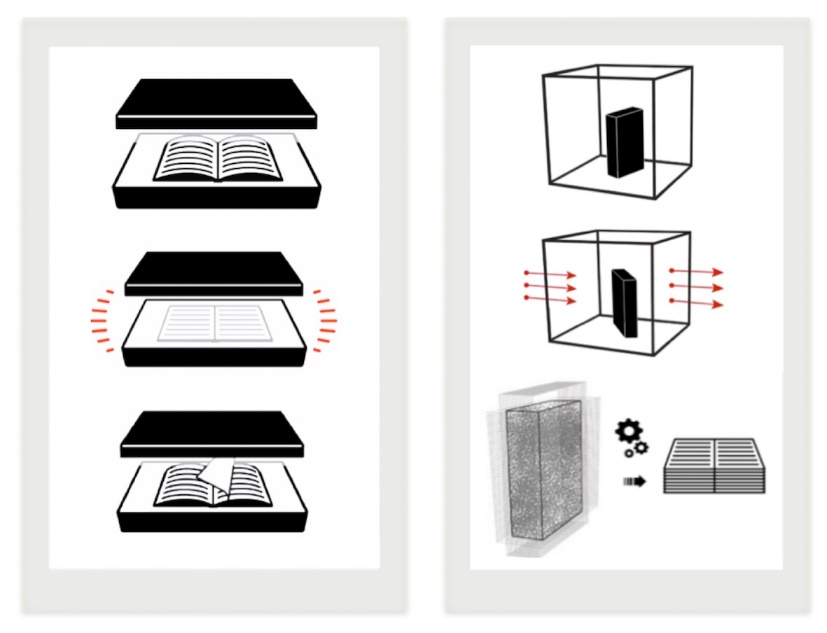
The use of X-ray tomography could facilitate and accelerate the first step of the digitization process: without opening the manuscripts, throughout the 3D X-ray imaging, the entire manuscript’s volume could be acquired and the information virtually extracted, page-by-page, from the 3D tomography volume (). This approach could make the reading of manuscripts possible without opening them in a completely non-invasive way: the technique strongly reduced the objects’ manipulation and eliminated the needing for page-turning, drastically reducing the risk of damages.
To explore this new approach, I carried out numerous tests and analyses, from the chemical investigation of the inks to the imaging with synchrotron source and conventional X-ray lab-based imaging systems. Here, I present the milestones achieved, from the first chemical investigation on a parchment fragment performed in 2014 to the tomographic imaging of a sealed 14th century Venetian last will acquired with one of the transportable X-ray set-up of the X-ray tomography for Cultural Heritage Laboratory of the Department of Physics and Astronomy Augusto Righi of the University of Bologna.
The approach of VXR is based on pioneering projects that exploited X-rays to decipher documents such as the use of synchrotron light to retrieve lost text from the Archimedes Palimpsest with X-ray fluorescence . V. Mocella pioneered the use of X-ray tomography to analyze handwriting in revealing letters in rolled Herculaneum papyri by X-ray phase-contrast imaging , and the works of W. Seales et al. and of G. R. Davis posed solid bases for the development of the tomographic technique for manuscripts. Furthermore, Patten et al. studied the potential document’s radiation damage.
Besides the studies I conducted in the framework of the Virtual X-ray reading project, and here presented , , , , , , , recently impressive result was obtained using X-ray tomography to virtually unroll a scroll from En-Gedi .
The success of these projects demonstrated the great potential of X-ray techniques in the study, deciphering, and reading of ancient manuscripts. Moreover, these studies open the way to using X-ray tomography to decipher texts on more variegated supports, such as the work by D. Stromer et al. and their research to the reading of a bamboo scroll [StromerBamboo2019] or the scan of a film footages by Liu et al. .
Traditional digitization vs. X-ray tomography digitization.
Along the path to reading manuscripts using X-ray tomography, a critical issue is the nature of the ancient inks. Before the advent of modern inks, two main groups of black inks were used for centuries: iron-based ones and, less common in Europe, carbon-based ones. X-ray absorption imaging is made possible for the first group by the heavy element content of the inks generically denominated iron gall - that corresponds to a wide variety of ingredients and recipes . On the contrary, X-ray absorption contrast is weak for carbon-based inks - although imaging could still be possible with alternate X-ray contrast mechanisms or with other imaging techniques.
Even if the use of iron-gall inks for centuries in Europe has been tested extensively , , , , , recipes and composition of the inks used for every day documents (as opposed to pieces of high artistic or historical value) and for archival documents - such as notary papers, work contracts, commercial transactions, and demographic accounts – is scarcely documented. Furthermore, Archives such as the Venetian Archivio di Stato collection span ten centuries, with inevitable fluctuations in the ink’s recipes.
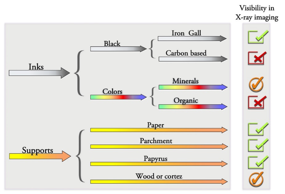
The major challenge of using X-ray tomography for digitization was – and still is – the virtual extraction of the manuscripts’ pages. The result of X-ray tomography is a 3D volume where each pixel in the 3D volume represents the X-ray absorption of the point of the real object. The identification, isolation, and extraction of the pixels related to the specific page are needed to extract the document’s foils. Several approaches have been used, most of them dedicated to papyrus tomographic imaging and their segmentation . In principle, a manual, supervised procedures or full unsupervised ad hoc algorithms are used to recognize and classify pages, numbering them and recognize the empty space between them. Then, only the classified pages will be extracted and rectified to be visualized. For these processes, the thickness of the pages, the spacing between them, and the resolution and quality of the tomographic image are crucial for a successful result. Unfortunately, the large variety of manuscripts – in forms, page number, and materials – make developing a generic and highly flexible algorithm a hard goal.
The VXR project analyzed all these aspects of the X-ray tomography digitization, from the chemical investigation of inks of every day documents to the feasibility study - with large facilities and laboratory instrumentations - of X-ray tomography to read and to digitize archival records and documents.
Ancient manuscripts’ materials
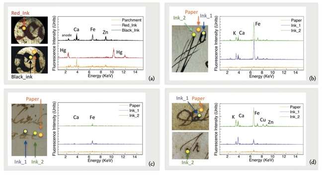
The X-ray visibility of writings in X-ray tomography – named the X-ray contrast – is induced by the difference in X-ray absorption between the support – the pages – and the writings.
Therefore, the chemistry of the inks plays a primary role: inks with a high content of heavy elements, such as iron and metals, are visible in X-ray images. Contraries’, inks with a low content of heavy elements or mainly composed of light elements, are almost invisible. Beside this, the supports and their thickness play an important part in the feasibility of the technique. In a schematization of the main manuscripts’ materials and their visibility in X-ray imaging is reported.
As mentioned, inks for writing could be divided into two main categories: black inks and colored ones. For many centuries, in Europe, the most widely used black ink for manuscripts was the iron gall, a name suggesting the presence of iron. Black inks with no heavy elements were less common, such as the Roman atramentum scriptorium, based on lampblack with a gum binder.
Although the generic iron gall formula corresponded to a wide variety of ingredients , the basic fabrication process was an acid reaction with an iron compound. The most common procedure involved tannic acid (C76H52O46) and iron sulfate (FeSO4) in rainwater, white wine, or vinegar . Tannic acid was obtained from plants, the richest source being the ‘galls’ produced by trees in response to parasite attacks (e.g. by gall wasps); for example, the British oak galls or the top-quality Aleppo galls. Iron sulfate was known as ‘green vitriol’, extracted from mines, notably coal mines. The reaction of tannic acid with FeSO4 produced, with oxygen exposure, ferrotannate, a black pigment. In addition to the pigment, the black inks also contained a water-soluble binder. One of the most common was gum arabic. This is a natural product of trees, e.g. acacia, rich in polysaccharides and glycoproteins and its main component is arabin , . Other ingredients could be present, such as logwood pigment. A dangerous property of iron gall inks is their corrosivity and their chemical attack to the substrate. The ink–substrate interaction was extensively analyzed by , , and using techniques such as synchrotron radiation XANES (X-ray absorption near-edge structure) and micro-fluorescence techniques.
In addition to black inks, for important writings or illuminated manuscripts, colored inks were used. Contrary to iron-gall inks, these inks are very similar to artist pigments. For them, the scientific literature is much vaster and the recipes remained unchanged for centuries, handed down from one generation into another, from one bottega to other. These aspects make these inks identification and study easier than for the black ones. Moreover, most of these pigments are mineral and contain elements visible in X-ray , making techniques such as MA-XRF the perfect approach to analyze them , , and X-ray tomography feasible in most cases.
As mentioned, supports are important for the visibility of the writings. Even if the X-ray absorption of almost all supports is similar and limited – cellulose or rag paper, parchment, wood – a higher manuscripts’ thickness decreased the total X-ray contrast of the writings. For this reason, almost all the supports could be digitized with X-ray tomography but a successful result is strictly connected with the manuscripts’ thickness.
Main manuscript materials and their visibility in X-ray
imaging.
A revolution in the world of texts in Europe appended with the invention the industrial printing by Johannes Gutenberg in 1455 , . The production of books became extremely rapid, exceptionally abundant in numbers, and increasingly less expensive than the traditional one . In few decades, an almost total conversion from handwritten to printed books occurred . Consequently, the materials also drastically changed: from the widespread use of the iron-gall inks for handwritten documents to the almost exclusive use of carbon-based inks for printing books. Moreover, the traditional variety of writing supports were replaced by mass-produced papers. Notably researches were carried out on the Gutenberg Bible inks , , , and the on elemental analysis of ancient papers by Manso et al. , , .
With the aim of investigates this extremely rapid change, in 2018 in collaboration with Cà Foscari University, we performed an extensive non-invasive investigation campaign of a large corpus of early printed books. We analyzed inks and papers of sixty volumes, part of the important collection of the Ateneo Veneto in Venice (Italy), printed between the 15th and the 17th centuries in the main European manufacturing centers .
X-ray analysis of inks
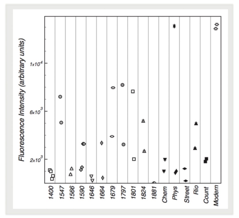
The ink chemistry of numerous manuscripts was analyzed using the X-ray Fluorescence (XRF), a completely non-invasive and non-destructive technique widely used to investigate Cultural Heritage objects , , . As detailed reported in , I analyzed more than 20 manuscripts’ - from a religious parchment to a 200-page physics book, from 16th century documents to a modern mock-up.
Different points were analyzed for each manuscript, including the support – parchment or paper – and several spots on inks. For the supports, XRF spectra confirm the light-element composition with no features that could affect our analysis. For inks analysis, the XRF ink spectra were corrected by subtracting those of the paper area. Although a full quantitative analysis is not possible, this correction and the use of the same acquisition conditions allowed a good comparison between data. In some examples of the XRF results on a 15th century parchment (a), and 1590 (b), 1646 (c), and 1664 (d) manuscripts. In inks, heavy or medium-heavy elements such as K, Ca, Fe, Cu, and Zn were found. For the 15th parchment, the analysis detected Hg in the red ink (a), most probably cinnabar.
The spectra evidence two important issues. Firstly, the ink composition varies from ink to ink – from an ink containing almost only a small amount of iron, as shown in c, to those containing several heavy elements such as the one of d. Moreover, the investigation revealed the high fluctuation in heavy-element contents, as illustrated in b-c.
To better investigate this aspect, I compared the iron content of all the analyzed manuscripts – as shown in . As explained in detail in , the investigation highlighted the fluctuation from manuscript to manuscript and does not show a clear time-related trend. Furthermore, the comparison shown high fluctuations from page to page of the same documents (1590 and Phys of ) and, more surprisingly, from words to words on the same manuscript pages (1679, 1797, 1801 of ). While the fluctuations between manuscripts, and maybe between pages, are most likely due to different inks, the latter effects could have been caused by the handwriting process itself, e.g., the variations of the rate of ink release by the pen.
These results pose one more challenge in the X-ray digitization approach: in the best situations, the ink iron content is high, and the X-ray attenuation image quality is good enough to perform the tomographic reconstruction of the writings. In the worst cases, even simple character detection is a problem. But the fluctuation inside the same manuscript does not guarantee a good visibility of the inks along with all the document.
XRF analysis of ink and paper of a 15th century parchment (a), 1590 (b, 1646 (c) and 1664 (d) manuscripts (Data from [9])
Iron content detected by XRF in seventeen manuscripts over six centuries. The last six specimens were written after the mid-18th century and Modern is a simulated stack written with iron gall ink (Data from [12])
To chemically study the early printing technology, to investigate its evolution and, potentially, to identify physical/chemical fingerprints of different manufactures and/or printing dates, we performed an extensive XRF campaign on sixty volumes, printed between the 15th and the 17th centuries in the main European manufacturing centers .
Differently for the XRF analysis of manuscripts, the nature of these inks – mainly carbon-based – and the low quantity of pigments left on the pages by the printing technique, made necessary the development of a more sophisticated data pre-treatments to analyze the paper and ink contribution independently.
As described in detail in , the analyses shown a high degree of homogeneity in all the materials analyzed. The XRF investigations detected the use of carbon-based inks for all the black features of the books, from the characters to the illustrations. Moreover, the ubiquitous presence of small amount of K, Ca, Fe and Mn and the frequent presence of Cu and Zn were found. The presence of elements such as K and Ca could be linked to the use of fillers, while for the other heavy elements contain could be hypothesized the use of different ink recipes , , , , or the transfer of particles from the metal types .
The papers’ analysis showed similar results, with the ubiquitous presence of small amount of K, Ca, Fe, and frequently of Mn. Here, the presence of K, Ca, and Fe could be directly linked to paper production: the K is linked to the Alum used as a mordant and Ca was used in all the phases of the paper production, from the gelatin size to the whitening agents , , , , , . Moreover, traces of metals in water are likely responsible for Fe, and Mn detection and an additional source could be identified in the particle transfer from the metal types during the printing process .
Even if the results confirm the ineffectiveness of X-ray tomography as a digitization technique for early printing books, the detailed statistical analyses identified potential fingerprints of different European manufacturers, particularly the Venetian ones .
X-ray imaging and tomography of ancient manuscripts
Imaging with synchrotron radiation
To investigate the feasibility of X-ray imaging on manuscripts, I analyzed different contrast mechanisms, i.e., absorption imaging and phase-related phenomena . I performed the firsts imaging experiments using a centralized synchrotron facility – the TOMCAT beamline of the Swiss Light Source (SLS). This source allows an extremely high spatial resolution (some microns), fast acquisition (some seconds), and imaging techniques with different contrast mechanisms such as free-propagation phase contrast and the differential phase contrast (DPC) .
As described in detail in , the first experiment concerned the tomography of a bundle of eight 0.8 cm-diameter fragments of a 1679 specimen stacked together to simulate a small volume. In the sample analyzed (a), the two three-dimensional reconstructed tomographic side views (b), and the comparison between the visible picture of the fragments and the tomographically reconstructed ‘pages’ (c). The exceptional spatial resolution (6.5 μm detector pixel) is largely sufficient not only to distinguish different ‘pages’ but also to determine whether the writing is on the front or the back of each ‘page’.
X-ray tomography with a synchrotron source of a 1679 manuscript’s
fragments (Data from [9])
To evaluate the capability of X-rays in detecting writings from different dates, the X-ray imaging of three manuscripts from 1590, 1664, and 1801 are acquired and shown in . As detailed described in , images were acquired at TOMCAT with the free-propagation operation mode and reflect both phase contrast and attenuation contrast. Here, phase contrast is primarily visible in the microscopic substrate features, while attenuation contrast prevails in the ink areas.
Besides the iron X-ray attenuation contrast visible in each document – that made the manuscript readable in X-rays - several other features almost invisible to the naked eye are identifiable. In all documents, the X-ray imaging makes visible the high fluctuation in iron content detected by XRF and proves that one of its causes is the ink released by the pen. Moreover, in imaging of 1664 manuscript (b) the signs of corrosion of the paper by the iron gall ink are much more evident than in naked eyes, and in c the traces of the papermaking process are perfectly visible as horizontal lines and emphasized by the phase-contrast mechanism.
Comparison between visible picture and X-ray imaging with a synchrotron
source of manuscripts from different epoch - 1590 (a&b), 1664 (c&d) and
1801 (e&d) (Data from [9])
In the case of low attenuation contrast, such as inks with a low iron content, the different imaging techniques provided by synchrotron light could be helpful. This is the case of the DPC imaging, where the acquisition process and the use of appropriate algorithms yield images corresponding to different contrast mechanisms: absorption, scattering, and refraction .
In the imaging results of the 15th century parchment acquired in DPC mode at TOMCAT . In b the DPC absorption-contrast image – equal to absorption radiography – with the excellent attenuation contrast of Hg-containing red characters, and the weak contrast of the black letters, in agreement with the low iron content detected by XRF (as shown in ).
c and d show scattering and refraction images. These seem to reflect the local specimen morphology (the parchment topography) rather than only it’s chemical composition. As described in , it is possible to argue that what we see in these figures corresponds to the substrate morphology, probably related to parchment damages or differences in its thickness (c), and to modification of the more superficial features, apparently related to the writing process or by the ink–substrate interaction. This could be used to detect a character when the attenuation contrast would only give a very faint picture.
Imaging with with different absorption methodology achieved with a
synchrotron source. 14 th parchment in visible light (a), absorption (b),
scattering (c) and refraction (d) (Data fro m [10] ).
X-ray imaging with lab equipment
The success of the tests performed at a centralized synchrotron facility demonstrates the approach feasibility but did not address two key issues: the needs of in situ analysis and the practicability of the technique on large areas manuscript. The first is crucial since moving specimens to such a facility creates conservative, logistic, practical, and even legal problems that would severely reduce the impact of the approach. The second is strictly related to the first ones since synchrotron emission is by its very nature highly collimated and typically used for small specimens. For these reasons, a laboratory X-ray imaging facility is imperative for the development of X-ray tomography digitization.
Initial imaging tests on large area manuscripts have been performed at the X-ray Imaging Center of the Pohang University of Science and Technology (POSTECH) in Korea . In the comparison of visible image and X-ray radiography of a single-page manuscript from 1679 acquired with laboratory equipment. The images show the excellent readability of the document in X-rays. Moreover, demonstrate the system’s capability in detecting the ink-content fluctuation and the effect of the intensity fluctuations due to the structures of the handmade paper and invisible in the conventional visible imaging. As described in detail in , several other successfully imaging tests have been performed, such as tomography of a stack of different pages of a modern manuscript created following ancient recipes – high-quality paper and iron-gall ink– and imaging of an ancient book rolled up to simulate a multi-page scroll.
All the tests proved the feasibility of the X-ray tomography technique to read manuscripts also with laboratory equipment, potentially available in situ and able to digitize large-areas manuscripts.
Comparison of visible image (a) and X-ray radiography (b) of a 1679
manuscript achieved with a lab-based source (Data fro m [11] ).
To evaluate the technique’s feasibility on large objects such as a large and thick manuscript, a large handwritten scientific manuscript (27 × 19 cm2) by an Italian scientist, Giulio Mancini, dated 1790–1800, was analyzed. The book consists of a 200-pages text with notes about different branches of physics and includes several inserts with drawings and graphs.
The book was analyzed with different imaging facilities: the μCT instrument available at the Center for X-ray Analytics at EMPA (Dübendorf, CH) and the tomographic set-up of the of X-ray Tomography for Cultural Heritage Laboratory of the University of Bologna .
The results shown in demonstrate the feasibility of the techniques with laboratory-based equipment also for large manuscripts. Thanks to the 3D tomographic volume, we can visualize the external part of the book (b), and we can navigate through all the 200 pages, where the writings are visible (c). Moreover, the word reading is feasible, and d shows several examples of clearly readably individual words from inside pages.
Nevertheless, the segmentation and extraction of the pages from these manuscripts are not trivial and pose severe challenges for the segmentation algorithms. As shown in c, the pages on the book’s fore-edge are almost unrecognizable, making the segmentation really arduous. The task is easier when significant air gaps exist between the pages and, in such cases, we can be optimistic about the feasibility of unsupervised page extraction algorithms. For samples such as this highly-compact book, the segmentation is extremely difficult, if not impossible with really powerful and sophisticated ad hoc algorithms.
X- ray tomography of a 200-pages book achieved with lab-based sources.
Visible picture and 3D image of the book (a), tomographic 3D image and view of the
inside pages (c), words and drawing extracted for the internal pages (d) (Data
from [12] and [13]).
In 2017, we performed a tomographic campaign of several closed and sealed last wills from the Venetian Archivio di Stato. The Archivio contains thousands of testaments that are still unopened: X-ray tomography constitutes the only chance to read them without breaking their official seals. In the first result of the campaign: the tomographic imaging of a 1351 Venetian testament, a paper document (about 5x10 cm2), still sealed and folded six times. The X-ray imaging were performed with the laboratory-based and transportable equipment of the X-ray Tomography Laboratory for Cultural Heritage of Bologna University.
As shown in b, the investigation detected words on the upper part of the external page and the strongly absorbing effect of the wax seal on the verso of the manuscript. To analyze all pages one-by-one, I performed a manual segmentation to separate the warped and closely-packed individual pages. In c, the individual segmented pages and, in d, parts of the texts and a complete internal page, extracted from the tomographic data.
Moreover, in X-ray tomography are readable the first lines of the external text: in d (left-top) the beginning of the document with a standard formula including the date, the place, and the name of the recording officer. This external portion of the text gives the opportunity to validate the tomography approach results, which reveals all pages equally well, internal and external.
The X-ray tomography made visible for the first time in six centuries part of the internal text and the first words of the document are perfectly readable: ”A nome de Dio” - ”In the name of God”, a standard formula of the beginning of last wills – d (left-middle). In d (left-bottom) many letters and several words from portions of internal pages are directly readable. In d (right) the reconstruction of the complete internal page, with lines of the text visible and few words directly readable and others inferable.
The noise effect that disturbs the text reading is caused by the characteristic inhomogeneity of the handmade paper. Also for this document are observable the contrast fluctuation from letter to letter and between different parts of individual letters. These fluctuations, combined with the noisy background, augment the difficulties of the virtual reading process. Moreover, it must be taken into account that the document is written in ancient Venetian, and most of the words are abbreviate - i.e. Venice was Rivoalti and written as Rti. This makes the reading of such documents more difficult and non-trivial for non-experts.
Tomographic investigation of a 15 th sealed Venetian document achieved
with the lab-based facility of UniBo. Visible pictures of the last will (a), 3D
tomographic volume (b) and segmented pages (c). In (d) part of the internal text
and a complete page (d) (Data from [13]).
Other imaging techniques
The VXR project proved the feasibility of the X-ray tomography and imaging of ancient manuscripts wrote with heavy-content inks, in particular for closed and sealed ones. But sometimes also the reading of open documents could be a challenge, such as in case of damaged manuscripts, faded or smudged writings, or overlapped texts. For these cases, the use of imaging techniques in the visible and near-visible light spectral range could be the key.
The visibility of inks and pigment is related to their absorption/reflectivity and these vary on the spectral region analyzed. The use of an extended spectral range offers several advantages, such as the increasing transparency of most of the pictorial materials as the wavelength increases (infrared reflectography), e.g. resulting in better visibility of the preparatory drawing of the miniatures thanks to the transparency of the overlaid pictorial layers. The use of the UV fluorescence imaging technique stimulates the fluorescence of the materials and makes it possible to emphasize details difficult to read – or hidden –in visible light. Moreover, preliminary considerations on materials’ identification could be made basing on their different appearance in the different spectral ranges.
Some examples of the behavior of inks in different regions of the spectra, part of the preliminary study of the sixty early printed books analyzed and presented in , are shown in , , and .
In the imaging of a front page of a 1525 Venetian printed book in visible light ( a), in the IR region (850 nm filter) b), and under UV light to perform UV fluorescence imaging c). The front page is written in red and black inks and two markers are present. While in IR imaging the black inks remain clearly readable, as well as the markers, the red one disappeared. The behavior of the black ink used for the printed text suggests the use of carbon-based ink. Indeed, while the iron-based inks are transparent to the IR radiation the carbon-based inks are strong IR absorbers . The high reflectivity of the red inks in the IR region could suggest the use of pigment such as cinnabar, minium, or red lake but the imaging technique used is not adequate for its identification.
The UV fluorescence imaging, stimulating the fluorescence of the paper, revealed inhomogeneities most probably related to humidity damages. Furthermore, a deeper observation of the markers revealed some smudges near them, probably due to some organic components.
Similar inks behavior were observed for the imaging of an illustrated page of a 1485 Venetian printed book shown in . Here as well, the pigments - yellow and red - became transparent in IR while the black text and illustration remain perfectly readable. Furthermore, the UV Fluorescence highlight evident humidity traces.
The imaging analysis of a 1499 Venetian printed book shown in put in evidence the behaviors of two different black inks: while the printed text and the illustration remain clearly visible in IR, the manuscript annotations disappear. Again, this suggests the use of carbon-based ink for the printed text and an iron-based one for handwriting .
Imaging in Visible Light (a), Infrared (b) and UV fluorescence (c) of a 1525 Venetian printed book. (Data from [18])
Imaging in Visible Light (a), IR (b) and UV fluorescence (c) of a 1485 Venetian printed book. (Data from [18])
Imaging in Visible Light (a), IR (b) and UV fluorescence (c) of a page of 1499 Venetian printed book. (Data from [15])
To analyze the inks used for early printed volumes, a XRF campaign was performed on the sixty book.
In , the XRF results of the inks of , and clarifies their nature. The analysis of the red ink used for the colored title of detects Hg and Pb, confirming the hypothesis made and suggesting the use of a mixture of Vermilion, and Red Lead. A similar composition is found for the red ink of the illustration of . The XRF of the yellow ink detected As, Pb and Sn, indicating a combination of Lead-Tin Yellow and Orpiment or Realgar. The investigation of the black inks of confirms the hypothesis of an iron-based ink for the handwritten annotation and a carbon-based one for the printed text.
XRF analysis of the ink of Figures 11,12 and 13 (Data from [18])
The multispectral images presented investigate wide spectral ranges, impeding to take full advantage of the different behaviors of the pigments in the different portions of the spectrum.
The use of techniques such as hyperspectral imaging allows exploiting these features to the maximum .
This technique consists of acquiring and measuring the reflectance spectra of the analyzed object in various wavelength ranges - usually in the range from 400 to 1000 nm in the visible and near-infrared regions and using 50–150 different spectral bands. The result is an image cube, made up of several images, each one corresponding to a particular wavelength band . The analysis and the reconstruction of the reflectance spectra of the pigments could allow their identification . Moreover, in the case of pigments similar in visible light, the acquisition of tens of monochromatic images enables their comparison in different parts of the spectrum and, potentially, distinguishes them. Similarly, in the case of superimposed images or writings, could be identified a spectral range in which the upper writing disappears, making the deeper readable.
Conclusions
The research and studies conducted in the Virtual X-ray Reading framework, from the extensive analysis of ancient inks to the radiographic and tomographic imaging of ancient documents using synchrotron and lab-based equipment, demonstrated the feasibility of the technique.
The iron-gall nature of the inks used for every-day manuscripts was tested by the extensive XRF analysis performed along with the project and partially reported here. Furthermore, the investigation highlighted one more challenge for X-ray imaging: the high fluctuation in heavy-element content of the writings could make the X-ray contrast inhomogeneous along with the texts. This, combined with the considerable inhomogeneity of the handmade paper, makes essential a strong image pre-processing to improve the readability of the texts.
The imaging feasibility tests, performed both with synchrotron facility and lab-based equipment, shown that the X-ray tomography of manuscripts and its reading is feasible, not only on small-size objects but also on books. Nevertheless, a robust segmentation methodology is needed, and its crucial for collection investigation, and it’s a formidable challenge.
Despite the challenges, the VXR project and its results proved that this approach is applicable to the vast majority of European manuscripts and, potentially, to collections worldwide.
Acknowledgements
I am grateful to Giorgio Margaritondo for his exceptional positive support and mentorship, Patrick Aebischer for his leadership of the VTM project, and the Verbantiqua bookstore (http://www.copernicum.it) for the generous donation of some of antique manuscript specimens here presented. Over the years the project involved several Professors, Researchers, and Laboratories and I am grateful to all of them. In particular, to the staff of the Swiss Light Source for their technical support, to Professor Ferruccio Petrucci, Eva Peccenini, and Anna Impallaria of the University of Ferrara for their assistance in the chemical analysis, to Yeukuang Hwu and Jung Ho Je for their support and for the analysis conducted at POSTEC, to the staff of EMPA for their technical support, to the Archivio di Stato in Venice and its staff for their precious collaboration, to the Ateneo Veneto for its essential help, to Dorit Raines for her extraordinary support and mentorship, and the X-ray tomography for Cultural Heritage Laboratory of the University of Bologna and its staff for the last years of working together.
The project was supported by the Ecole Polytechnique Fédérale de Lausanne (EPFL) under the general framework of the Venice Time Machine Project, supported by the Lombard Odier Foundation. Over the years, the researches were supported by various organizations: by the Fonds National Suisse pour la Recherche Scientifique, by the Center for Biomedical Imaging (CIBM), by the National Research Foundation of Korea (NRF), by the National Science and Technology Program for Nanoscience and Nanotechnology, Taiwan, by the Thematic Research Project of Academia Sinica (Taiwan) and by the MIUR National Access Pilot call (IPERIONCH.it, IPERION CH Grant Agreement n. 654028).
References
Kaplan, Frederic, How to build an information time machine: Venice time machine. Technical report, EPFL, Digital Humanities Lab, 2013. http:// dhvenice.eu/
Bergmann, Uwe, X-ray fluorescence imaging of the Archimedes Palimpsest: a technical summary. Technical Summary, Report 100, SLAC, 2000
Mocella, Vito, Emmanuel Brun, Claudio Ferrero, and Daniel Delattre. Revealing Letters in Rolled Herculaneum Papyri by X-Ray Phase-Contrast Imaging. Nature Communications 6, no. 1 (January 20, 2015). https://doi.org/10.1038/ncomms6895.
Lin, Yun and Seales, William, Brent, Proceedings of the 10th IEEE International Conference on Computer Vision, Vol. 1, pp. 662–669. (2005).
Baumann, Rayan, Porter, Dorothy Carr and Seales, William, Brent, The use of micro-CT in the study of archaeological artifacts, Proceedings of 9th Int Conf on NDT of Art (2008).
Seales, William, Brent., Griffioen, J., Baumann, R. & Field, M. (2011). 10th International Conference on Non-Destructive Investigations and Microanalysis for the Diagnostics and Conservation of Cultural and Environmental Heritage, pp. 1–9. Brescia: AIPnD.
Mills, David, Oksana Samko, Paul Rosin, Kate Thomas, Tim Wess, and Graham R. Davis. Apocalypto: Revealing the Unreadable. In Developments in X-Ray Tomography VIII, edited by Stuart R. Stock. SPIE, 2012. https://doi.org/10.1117/12.928917..
Patten, Kate, Lee Gonzalez, Craig Kennedy, David Mills, Graham Davis, and Tim Wess. Is There Evidence for Change to Collagen within Parchment Samples after Exposure to an X-Ray Dose during High Contrast X-Ray Microtomography? A Multi Technique Investigation. Heritage Science 1, no. 1 (2013): 22. https://doi.org/10.1186/2050-7445-1-22.
Albertin, Fauzia, Astolfo, Alberto, Stampanoni, Marco, Peccenini, Eva, Hwu, Yeukuang, Kaplan Frederic and Margaritondo Giorgio. Ancient Administrative Handwritten Documents: X-Ray Analysis and Imaging. Journal of Synchrotron Radiation 22, no. 2 (January 30, 2015): 446–51. https://doi.org/10.1107/s1600577515000314.
Albertin, Fauzia, Astolfo, Alberto Stampanoni, Marco, Peccenini, Eva, Hwu, Yeukuang, Kaplan, Frederic and Margaritondo Giorgio. X-Ray Spectrometry and Imaging for Ancient Administrative Handwritten Documents. X-Ray Spectrometry 44, no. 3 (January 13, 2015): 93–98. https://doi.org/10.1002/xrs.2581.
Albertin, Fauzia, Peccenini, Eva, Hwu, Yeukuang, Lee, Tsung-Tse, Ong, E.B.L. , Je, Jung Ho, Kaplan, Frederic and Margaritondo Giorgio, The Venice ” Archivio Di Stato ”: Innovating Digitization With X-Ray Tomography, in: Proc. 2015 Digit. Herit. Congr., 2015: pp. 6–11.
Albertin, Fauzia, Patera, Alessandra, Jerjen, Ian, Hartmann, Stephan, Peccenini, Eva, Kaplan, Frederic, Stampanoni, Marco, Kaufmann, Rolf and Margaritondo, Giorgio. Virtual Reading of a Large Ancient Handwritten Science Book. Microchemical Journal 125 (March 2016): 185–89. https://doi.org/10.1016/j.microc.2015.11.024.
Albertin, Fauzia, Romito, Marilisa, Peccenini, Eva, Bettuzzi, Matteo, Brancaccio, Rosa, Morigi, Maria Pia, del Rio, Monica, Raines, Dorit, Margaritondo, Giorgio and Psaltis Demetri. From Closed Testaments to Books: Virtual X-Ray Reading as an Alternate Digitization Technology for Fragile Documents. Archiving Conference 2017, no. 1 (May 15, 2017): 14–18. https://doi.org/10.2352/issn.2168-3204.2017.1.0.14.
Bettuzzi, Matteo, Albertin, Fauzia, Brancaccio, Rosa, Casali, Franco, Morigi, Maria Pia, Peccenini, Eva, X-Ray Computed Tomography Applied to Investigate Ancient Manuscripts. JB. Il Nuovo Cimento C 40, no. 2 (September 5, 2017): 1–9. https://doi.org/10.1393/ncc/i2017-17102-x.
Albertin, Fauzia, Balliana, Eleonora, Pizzol, G., Colavizza, Giovanni, Zendri, Eleonora and Raines, Dorit, “Printing Materials and Technologies in the 15th–17th Century Book Production: An Undervalued Research Field.” Microchemical Journal 138 (May 2018): 147–53. https://doi.org/10.1016/j.microc.2017.12.010.
Seales, William Brent, Clifford Seth Parker, Michael Segal, Emanuel Tov, Pnina Shor, and Yosef Porath. From Damage to Discovery via Virtual Unwrapping: Reading the Scroll from En-Gedi. Science Advances 2, no. 9 (September 2016): e1601247. https://doi.org/10.1126/sciadv.1601247.
Stromer, Daniel, Vincent Christlein, Christine Martindale, Patrick Zippert, Eric Haltenberger, Tino Hausotte, and Andreas Maier. Browsing through Sealed Historical Manuscripts by Using 3-D Computed Tomography with Low-Brilliance X-Ray Sources. Scientific Reports 8, no. 1 (October 18, 2018). https://doi.org/10.1038/s41598-018-33685-4.
Stromer, Daniel, Vincent Christlein, Xiaolin Huang, Patrick Zippert, Tino Hausotte, and Andreas Maier. Virtual Cleaning and Unwrapping of Non-Invasively Digitized Soiled Bamboo Scrolls. Scientific Reports 9, no. 1 (February 19, 2019). https://doi.org/10.1038/s41598-019-39447-0
Liu, Chang, Paul L. Rosin, Yu-Kun Lai, Graham R. Davis, David Mills, and Charles Norton. Recovering Historical Film Footage by Processing Microtomographic Images. In Digital Heritage. Progress in Cultural Heritage: Documentation, Preservation, and Protection, 219–31. Springer International Publishing, 2016. https://doi.org/10.1007/978-3-319-48496-9_18
Yale University Library Special Collections Conservator Unit, Preservation Department. Medieval Manuscripts, Some Ink and Pigment Recipes. Yale University, 2012.
Carmine, P. D., Giuntini, L., Hooper, W., Lucarelli, F., Mandó, P., Further results from PIXE analysis of inks in Galileo’s notes on motion, Nucl. Inst. Methods B 113 (1996) 354–356.
Lucarelli, F., Mandó, P., Recent applications to the study of ancient inks with external-PIXE facility, Nucl. Inst. Methods B 109 (1996) 644–652.
Hahn, O., W. Malzer, B. Kanngiesser, and B. Beckhoff. Characterization of Iron-Gall Inks in Historical Manuscripts and Music Compositions Using x-Ray Fluorescence Spectrometry. X-Ray Spectrometry 33, no. 4 (July 2004): 234–39. https://doi.org/10.1002/xrs.677
Boadas, Sònia. Techniques and Instruments for Studying the Autograph Manuscripts of Lope de Vega. Hipogrifo. Revista de Literatura y Cultura Del Siglo de Oro 8, no. 2 (December 2020): 509–31. https://doi.org/10.13035/h.2020.08.02.30.
Stabile, Sara, Francesca Palermo, Inna Bukreeva, Daniela Mele, Vincenzo Formoso, Roberto Bartolino, and Alessia Cedola. A Computational Platform for the Virtual Unfolding of Herculaneum Papyri. Scientific Reports 11, no. 1 (January 18, 2021). https://doi.org/10.1038/s41598-020-80458-z.
Liu, Chang, Paul L. Rosin, Yu-Kun Lai, and Weiduo Hu. Robust Virtual Unrolling of Historical Parchment XMT Images. IEEE Transactions on Image Processing 27, no. 4 (April 2018): 1914–26. https://doi.org/10.1109/tip.2017.2783626.
Bukreeva, I., A. Mittone, A. Bravin, G. Festa, M. Alessandrelli, P. Coan, V. Formoso, et al. Virtual Unrolling and Deciphering of Herculaneum Papyri by X-Ray Phase-Contrast Tomography. Scientific Reports 6, no. 1 (June 6, 2016). https://doi.org/10.1038/srep27227.
Stromer, Daniel, Vincent Christlein, Tobias Schoen, Wolfgang Holub, and Andreas Maier. Browsing through Closed Books: Fully Automatic Book Page Extraction from a 3-D X-Ray CT Volume. In 2017 14th IAPR International Conference on Document Analysis and Recognition (ICDAR). IEEE, 2017. https://doi.org/10.1109/icdar.2017.45.
Smith, G. (2009). Chem. Ed. NZ, May 2009 Issue, pp. 12–15.
Ruggiero, D. (2002). Report for Laboratorio di Fisica dell’Istituto per il Restauro e la Conservazione del Patrimonio Archivistico e Librario, pp. 1–32.
Banik, G., Mairinger, F. & Stachelberger, H. (1983). Restaurator, 5, 71–93.
Neevel, J. G. & Reissland, B. (1997). Proceedings of the Workshop on Iron Gall Ink Corrosion, pp. 37–45. Amsterdam: Netherlands Institute of Cultural Heritage.
Proost, K., Janssens, K., Wagner, B., Bulska, E. & Schreiner, M. (2004). Nucl. Instrum. Methods Phys. Res. B, 213, 723–728.
Kanngiesser, B., Hahn, O., Wilke, M., Nekat, B., Malzer, W. & Erko,A. (2004). Spectrochim. Acta B, 59, 1511–1516.
Gettens, R. J. et. al., Artist’s pigments: a handbook of their history and characteristics, Cambridge University Press and National Gallery of Art, 1993
Delaney, John K., Paola Ricciardi, Lisha Deming Glinsman, Michelle Facini, Mathieu Thoury, Michael Palmer, and E. René de la Rie. Use of Imaging Spectroscopy, Fiber Optic Reflectance Spectroscopy, and X-Ray Fluorescence to Map and Identify Pigments in Illuminated Manuscripts. Studies in Conservation 59, no. 2 (January 25, 2014): 91–101. https://doi.org/10.1179/2047058412y.0000000078..
Mazzinghi, Anna, Chiara Ruberto, Lisa Castelli, Paola Ricciardi, Caroline Czelusniak, Lorenzo Giuntini, Pier Andrea Mandò, Marco Manetti, Lara Palla, and Francesco Taccetti. The Importance of Being Little: MA‐XRF on Manuscripts on a Venetian Island. X-Ray Spectrometry 50, no. 4 (July 16, 2020): 272–78. https://doi.org/10.1002/xrs.3181
Legrand, Stijn, Paola Ricciardi, Luca Nodari, and Koen Janssens. Non-Invasive Analysis of a 15th Century Illuminated Manuscript Fragment: Point-Based vs Imaging Spectroscopy. Microchemical Journal 138 (May 2018): 162–72. https://doi.org/10.1016/j.microc.2018.01.001.
Ricciardi, Paola, Stijn Legrand, Giulia Bertolotti, and Koen Janssens. Macro X-Ray Fluorescence (MA-XRF) Scanning of Illuminated Manuscript Fragments: Potentialities and Challenges. Microchemical Journal 124 (January 2016): 785–91. https://doi.org/10.1016/j.microc.2015.10.020.
Kapr, A., Johann Gutenberg: The Man and His Invention, Scolar Press, Aldershot, Hants, 1996.
Eliot, Simon, and Jonathan Rose, eds. A Companion to the History of the Book. Blackwell Publishing Ltd, 2007. https://doi.org/10.1002/9780470690949.
Eisenstein, E. L., The Printing Press as an Agent of Change, Cambridge University Press. 1980.
Febvre, L., Martin, H.-J., The Coming of the Book: The Impact of Printing 1450–1800, Verso, London, 1997
Schwab, R.N., Cahill, T.A., Kusko, B.H., Wick, D.L., Cyclotron analysis of the ink in the 42-line Bible, Pap. Bibliogr. Soc. Am. 77 (1983) 285–315.
Schwab, R.N., Cahill, T.A., Eldred, R.A., Kusko, B.H., Wick, D.L., New Evidence on the Printing of the Gutenberg Bible: The Inks in the Doheny Copy, Pap. Bibliogr. Soc. Am. 79 (1985) 375–410.
Needham, P., Division of Copy in the Gutenberg Bible: Three Glosses on the Ink Evidence, Pap. Bibliogr. Soc. Am. 79 (1985) 411–426.
Kusko, B.H., Cahill, T.A., Eldred, R.A. , Schwab, R.N. , Proton milliprobe analyses of the Gutenberg Bible, Nucl. Inst. Methods Phys. Res. B (1984) 689–694.
Manso, M., M. Costa, and M.L. Carvalho. X-Ray Fluorescence Spectrometry on Paper Characterization: A Case Study on XVIII and XIX Century Documents. Spectrochimica Acta Part B: Atomic Spectroscopy 63, no. 11 (November 2008): 1320–23. https://doi.org/10.1016/j.sab.2008.07.001.
Manso, M., M. Costa, and M.L. Carvalho. Comparison of Elemental Content on Modern and Ancient Papers by EDXRF. Applied Physics A 90, no. 1 (September 1, 2007): 43–48. https://doi.org/10.1007/s00339-007-4235-y.
Manso, M., and M. L. Carvalho. Elemental Identification of Document Paper by X-Ray Fluorescence Spectrometry. J. Anal. At. Spectrom. 22, no. 2 (2007): 164–70. https://doi.org/10.1039/b608361g.
Albertin, Fauzia, Chiara Ruberto, Costanza Cucci, Marco Callieri, Marco Potenziani, Eliana Siotto, Paolo Pingi, et al. ‘Ecce Homo’ by Antonello Da Messina, from Non-Invasive Investigations to Data Fusion and Dissemination. Scientific Reports 11, no. 1 (August 5, 2021). https://doi.org/10.1038/s41598-021-95212-2.
C. Dupont, Gelatine sizing of Paper and its Impact on the Degradation of Cellulose During Agening: A Study Using Size-exclusion Chromatography, Ph.D. thesis. University of Amsterdam. 2003.
T. Barrett, M. Ormsby, J.B. Lang, Non-destructive analysis of 14th–19th century European handmade papers, Restaurator 37 (2016) 93–135.
Missori, Mauro, Marcofabio Righini, and Anne-Laurence Dupont. Gelatine Sizing and Discoloration: A Comparative Study of Optical Spectra Obtained from Ancient and Artificially Aged Modern Papers. Optics Communications 263, no. 2 (July 2006): 289–94. https://doi.org/10.1016/j.optcom.2006.02.004.
Margaritondo, G. & Hwu, Y. (2013). Nuovo. Saggiatore, 29, 45–58.
Stampanoni, M., A. Groso, A. Isenegger, G. Mikuljan, Q. Chen, D. Meister, M. Lange, R. Betemps, S. Henein, and R. Abela. TOMCAT: A Beamline for TOmographic Microscopy and Coherent RAdiology ExperimenTs. In AIP Conference Proceedings. AIP, 2007. https://doi.org/10.1063/1.2436193.
Weitkamp, Timm, Ana Diaz, Christian David, Franz Pfeiffer, Marco Stampanoni, Peter Cloetens, and Eric Ziegler. X-Ray Phase Imaging with a Grating Interferometer. Optics Express 13, no. 16 (August 8, 2005): 6296. https://doi.org/10.1364/opex.13.006296.
R. Padoan, T. Steemers, M. Klein, B. Aalderink, QuantitativeHyperspectral Imag- ing of Historical Documents: Technique and Application, 9th International Conference on NDT of Art, Jerusalem Israel, 25-30 May 2008, 2008, pp. 1–10.
Hayem-Ghez, Anita, Elisabeth Ravaud, Clotilde Boust, Gilles Bastian, Michel Menu, and Nancy Brodie-Linder. Characterizing Pigments with Hyperspectral Imaging Variable False-Color Composites. Applied Physics A 121, no. 3 (September 8, 2015): 939–47. https://doi.org/10.1007/s00339-015-9458-8.
Striova, J., A. Dal Fovo, and R. Fontana. Reflectance Imaging Spectroscopy in Heritage. La Rivista Del Nuovo Cimento 43, no. 10 (October 2020): 515–66. https://doi.org/10.1007/s40766-020-00011-6.
![X-ray tomography with a synchrotron source of a 1679 manuscript’s fragments (Data from [9])](https://umanisticadigitale.unibo.it/article/download/14399/version/12724/14068/55557/image5.jpeg)
![Comparison between visible picture and X-ray imaging with a synchrotron source of manuscripts from different epoch - 1590 (a&b), 1664 (c&d) and 1801 (e&d) (Data from [9]).](https://umanisticadigitale.unibo.it/article/download/14399/version/12724/14068/55558/image6.jpeg)
![Imaging with with different absorption methodology achieved with a synchrotron source. 14 th parchment in visible light (a), absorption (b), scattering (c) and refraction (d) (Data fro m [10] ).](https://umanisticadigitale.unibo.it/article/download/14399/version/12724/14068/55559/image7.jpeg)
![Comparison of visible image (a) and X-ray radiography (b) of a 1679 manuscript achieved with a lab-based source (Data fro m [11] ).](https://umanisticadigitale.unibo.it/article/download/14399/version/12724/14068/55560/image8.jpeg)
![X- ray tomography of a 200-pages book achieved with lab-based sources. Visible picture and 3D image of the book (a), tomographic 3D image and view of the inside pages (c), words and drawing extracted for the internal pages (d) (Data from [12] an d [13] ).](https://umanisticadigitale.unibo.it/article/download/14399/version/12724/14068/55561/image9.jpeg)
![Tomographic investigation of a 15 th sealed Venetian document achieved with the lab-based facility of UniBo. Visible pictures of the last will (a), 3D tomographic volume (b) and segmented pages (c). In (d) part of the internal text and a complete page (d) (Data fro m [13] ).](https://umanisticadigitale.unibo.it/article/download/14399/version/12724/14068/55562/image10.jpeg)
![Imaging in Visible Light (a), Infrared (b) and UV fluorescence (c) of a 1525 Venetian printed book. (Data from [18])](https://umanisticadigitale.unibo.it/article/download/14399/version/12724/14068/56832/image11.jpeg)
![Imaging in Visible Light (a), IR (b) and UV fluorescence (c) of a 1485 Venetian printed book. (Data from [18])](https://umanisticadigitale.unibo.it/article/download/14399/version/12724/14068/56833/image12.jpeg)
![Imaging in Visible Light (a), IR (b) and UV fluorescence (c) of a page of 1499 Venetian printed book. (Data from [15])](https://umanisticadigitale.unibo.it/article/download/14399/version/12724/14068/56834/image13.jpeg)
![XRF analysis of the ink of Figures 11,12 and 13 (Data from [18])](https://umanisticadigitale.unibo.it/article/download/14399/version/12724/14068/56835/image14.jpeg)